Anatomy Of Chest Muscles / Chest Muscles How To Use The Chest Anatomy For Better Results / (1) the pectoralis major, and (2) the pectoralis minor.. Anatomically, the axilla is taken to have an apex, a base and four walls, three of which formed by muscles (snell, 1999; And flexibility to aid in the functional process of respiration. Each of these muscles has its origin on the scapula and inserts around the head of the humerus. Lateral lip of intertubercular groove of the. Clavicular head & sternocostal head.
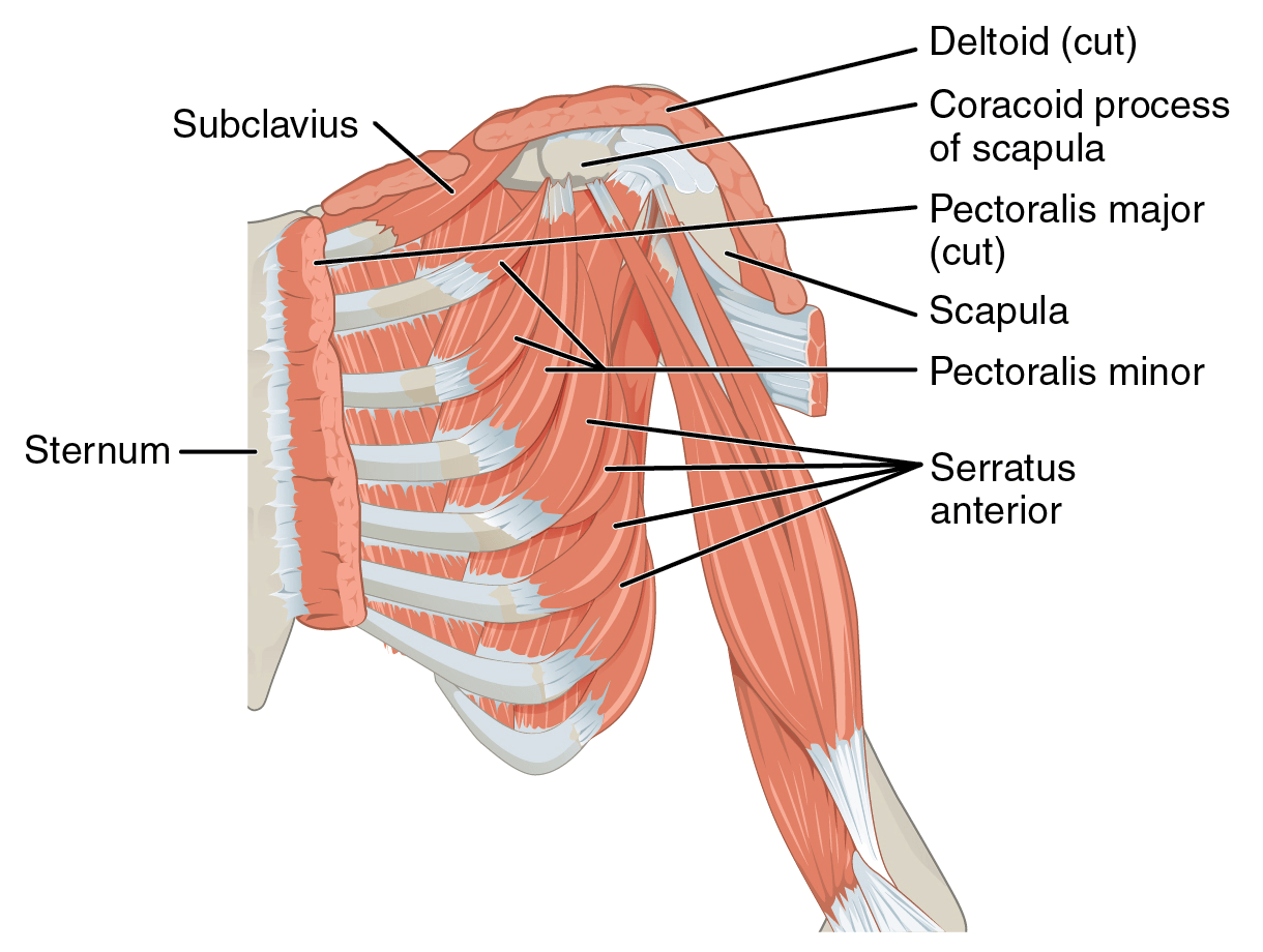
Anatomically, the axilla is taken to have an apex, a base and four walls, three of which formed by muscles (snell, 1999; Each of these muscles has its origin on the scapula and inserts around the head of the humerus. This provides an overview of the chest muscle group. The chest anatomy includes the pectoralis major, pectoralis minor, and the serratus anterior. Learn about each of these muscles, their locations, functional anatomy and exercises for them.
How To Release The Pectoralis Minor Ms Physiotherapy from msphysiotherapyaustralia.com Each of these muscles has its origin on the scapula and inserts around the head of the humerus. This divides the chest into two parts. Related posts of chest muscles diagram muscle anatomy in thigh. Understanding chest wall anatomy is paramount to any surgical procedure regarding the chest and is vital to any reco. The pectoralis major and the pectoralis minor, known collectively as your pecs. Muscle anatomy in thigh 12 photos of the muscle anatomy in thigh muscle anatomy inner thigh, muscle anatomy of thigh, muscle anatomy of upper thigh, muscle anatomy posterior thigh, muscle anatomy thigh mri, human muscles, muscle anatomy inner thigh, muscle anatomy of thigh, muscle anatomy of upper thigh, muscle anatomy. The pectoralis major muscles (also known as the pecs) are located on the front of the rib cage. The chest wall is a complex system that provides rigid protection to the vital organs such as the heart, lungs, and liver;
It's considered to be one of the most effective and reliable methods of measuring muscle activity.
22.06.2015 · chest muscles anatomy the chest is made up primarily of two muscles: Supraspinatus, infraspinatus, subscapularis, and teres minor. The muscles of the chest and upper back occupy the thoracic region of the body inferior to the neck and superior to the abdominal region and include the muscles of the shoulders. Stability to arm and shoulder movement; As a 15 year old female, you're still undergoing anatomical and physiological as a male who use to think that he was born a trans as he had a lot of chest fat (fact was i had around almost 40% body fat by the time i was. Rollover image shows the location of the major muscles of the chest, such as the pectoralis major, rectus abdominis, and external oblique. Use the mouse scroll wheel to move the images up and down alternatively use the tiny arrows (>>) on both side of the image to move the images.>>) on both side of the image to move the images. The tendons of these muscles surround and support the humerus while the contraction of the muscles rotates, adducts, or abducts the humerus. The beginner as well as advanced players meet with the problem when building a powerful chest. This webpage presents the anatomical structures found on thigh mri. Anatomy chest muscles illustrations & vectors. Beneath the pectoralis major is the pectoralis minor, a thin, triangular muscle. Muscle anatomy in thigh 12 photos of the muscle anatomy in thigh muscle anatomy inner thigh, muscle anatomy of thigh, muscle anatomy of upper thigh, muscle anatomy posterior thigh, muscle anatomy thigh mri, human muscles, muscle anatomy inner thigh, muscle anatomy of thigh, muscle anatomy of upper thigh, muscle anatomy.
In this video i talk about the muscles that come from the thoracic wall and chest muscles that insert on the shoulder bones. Muscles the dominant muscle in the upper chest is the pectoralis major. Sternocleidomastoid muscle clavicle and ribs anatomy muscle anatomy chest sternocleidomastoid ribs anatomy chest muscles anatomy thorax rib muscles chest muscles chest anatomy illustration. The pectoralis major muscles (also known as the pecs) are located on the front of the rib cage. Learn about each of these muscles, their locations, functional anatomy, and exercises for them.
3d Chest Human Anatomy Or Anatomical And Muscle Set Or Collection Stock Illustration Illustration Of Musculature Collection 200038397 from thumbs.dreamstime.com Sternocleidomastoid muscle clavicle and ribs anatomy muscle anatomy chest sternocleidomastoid ribs anatomy chest muscles anatomy thorax rib muscles chest muscles chest anatomy illustration. All about the chest muscles the chest anatomy includes the pectoralis major, pectoralis minor and the serratus anterior. These include pectoralis major, pectoralis minor, serratus anterior, and subclavius. The coracobrachialis and pectoralis major muscles connect the humerus anteriorly to the scapula and ribs, flexing and adducting the arm toward the front of the body when you reach forward to grab an object. The rotator cuff consists of four muscles: This webpage presents the anatomical structures found on thigh mri. Anatomically, the axilla is taken to have an apex, a base and four walls, three of which formed by muscles (snell, 1999; Browse 2,552 female chest anatomy stock photos and images available, or start a new search to explore more stock photos and images.
The pectoralis major, pectoralis minor, serratus anterior and subclavius.
Anatomy of chest muscles female.muscle basics and cellular components, naming of the muscles, and cat. This mri chest (thorax) axial cross sectional anatomy tool is absolutely free to use. Nine muscles of the chest and upper back are used to move the humerus (upper arm bone). Four main muscles in the pectoral region exert a force on the upper limb. Anatomy chest muscles illustrations & vectors. The chest wall is a complex system that provides rigid protection to the vital organs such as the heart, lungs, and liver; The rotator cuff consists of four muscles: The beginner as well as advanced players meet with the problem when building a powerful chest. Muscle anatomy neck 12 photos of the muscle anatomy neck dog neck muscle anatomy, front neck muscle anatomy, muscle anatomy neck, muscle anatomy of neck and shoulder, neck muscle anatomy chart, human muscles, dog neck muscle anatomy, front neck muscle anatomy, muscle anatomy neck, muscle. The chest or thorax is the region between the neck and diaphragm that encloses organs, such as the heart, lungs, esophagus, trachea, and thoracic diaphragm. Here, we break down the anatomy of your chest muscles. 22.06.2015 · chest muscles anatomy the chest is made up primarily of two muscles: It's considered to be one of the most effective and reliable methods of measuring muscle activity.
Function of the chest muscles Let's have a detailed look at each of their types and functions. And flexibility to aid in the functional process of respiration. Supraspinatus, infraspinatus, subscapularis, and teres minor. Plus, how to target each to make them bigger and stronger.
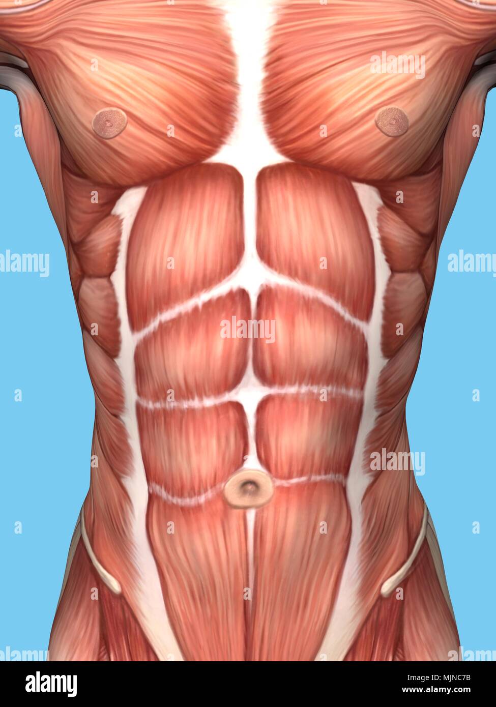
Anatomy Of Male Chest And Torso Featuring Major Muscular Groups Stock Photo Alamy from c8.alamy.com As a 15 year old female, you're still undergoing anatomical and physiological as a male who use to think that he was born a trans as he had a lot of chest fat (fact was i had around almost 40% body fat by the time i was. Anatomy chest muscles illustrations & vectors. This webpage presents the anatomical structures found on thigh mri. The pectoral region is located on the anterior chest wall. Muscles the dominant muscle in the upper chest is the pectoralis major. And flexibility to aid in the functional process of respiration. The pectoralis major and the pectoralis minor, known collectively as your pecs. It contains four muscles that exert a force on the upper limb:
The pectoralis major muscles (also known as the pecs) are located on the front of the rib cage.
Plus, how to target each to make them bigger and stronger. They are the pectoralis major, pectoralis minor, and the serratus anterior. The chest anatomy includes the pectoralis major, pectoralis minor, and the serratus anterior. These include pectoralis major, pectoralis minor, serratus anterior, and subclavius. As a 15 year old female, you're still undergoing anatomical and physiological as a male who use to think that he was born a trans as he had a lot of chest fat (fact was i had around almost 40% body fat by the time i was. Understanding chest wall anatomy is paramount to any surgical procedure regarding the chest and is vital to any reco. Supraspinatus, infraspinatus, subscapularis, and teres minor. Here, we break down the anatomy of your chest muscles. Use the mouse scroll wheel to move the images up and down alternatively use the tiny arrows (>>) on both side of the image to move the images.>>) on both side of the image to move the images. The chest or thorax is the region between the neck and diaphragm that encloses organs, such as the heart, lungs, esophagus, trachea, and thoracic diaphragm. Muscles the dominant muscle in the upper chest is the pectoralis major. Sternocleidomastoid muscle clavicle and ribs anatomy muscle anatomy chest sternocleidomastoid ribs anatomy chest muscles anatomy thorax rib muscles chest muscles chest anatomy illustration. To know whether or not an exercise targets the right muscles or not, scientists use a type of test called electromyography (emg).
Computed tomography (ct) of the chest can detect pathology that may not show up on a conventional chest radiograph(1) anatomy of chest. To know whether or not an exercise targets the right muscles or not, scientists use a type of test called electromyography (emg).


0 Komentar